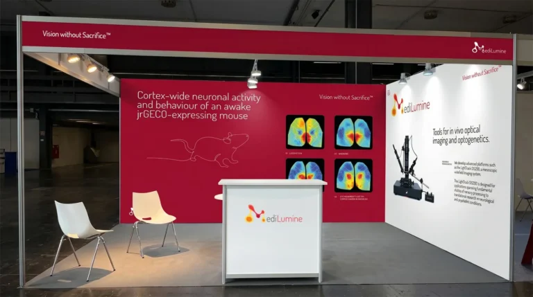
MediLumine at FENS 2026: Visit Us at Booth 159
We’re excited to be exhibiting at FENS 2026 in Barcelona, and we’d love to see you at Booth 159. Stop by to explore our full


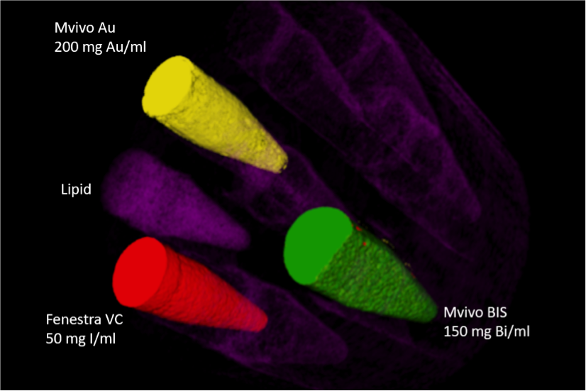
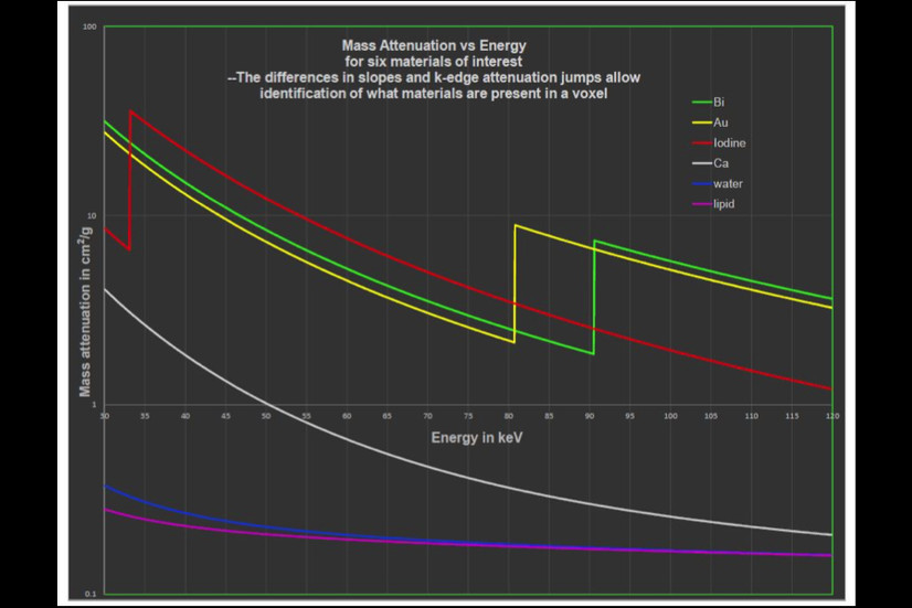
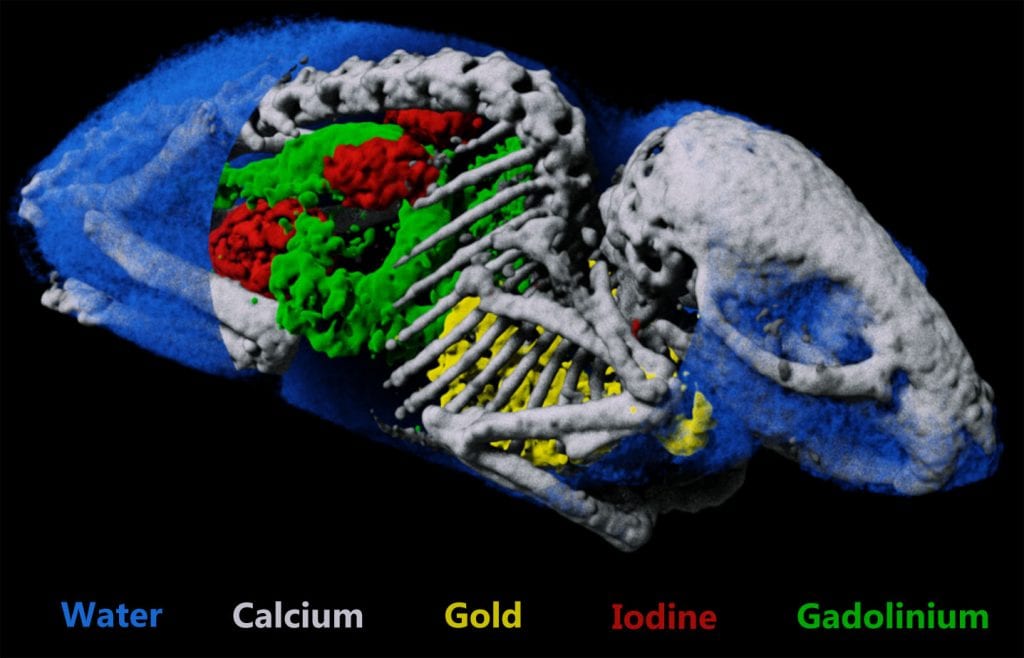
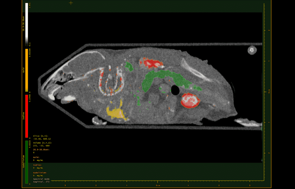
I'm really excited about this partnership as it combines the power of MARS quantitative imaging with the MediLumine's functional contrast agents. The combination of the two technologies greatly increases the opportunity for novel molecular imaging for pre-clinical researchers.

Working with MARS Bioimaging unlocks the potential of color x-ray imaging by providing a means to visualize disease in ways not yet imagined. Contrast enhanced color x-ray imaging will become a powerful tool in understanding human diseases and advancing drug discovery programs internationally.

We’re excited to be exhibiting at FENS 2026 in Barcelona, and we’d love to see you at Booth 159. Stop by to explore our full
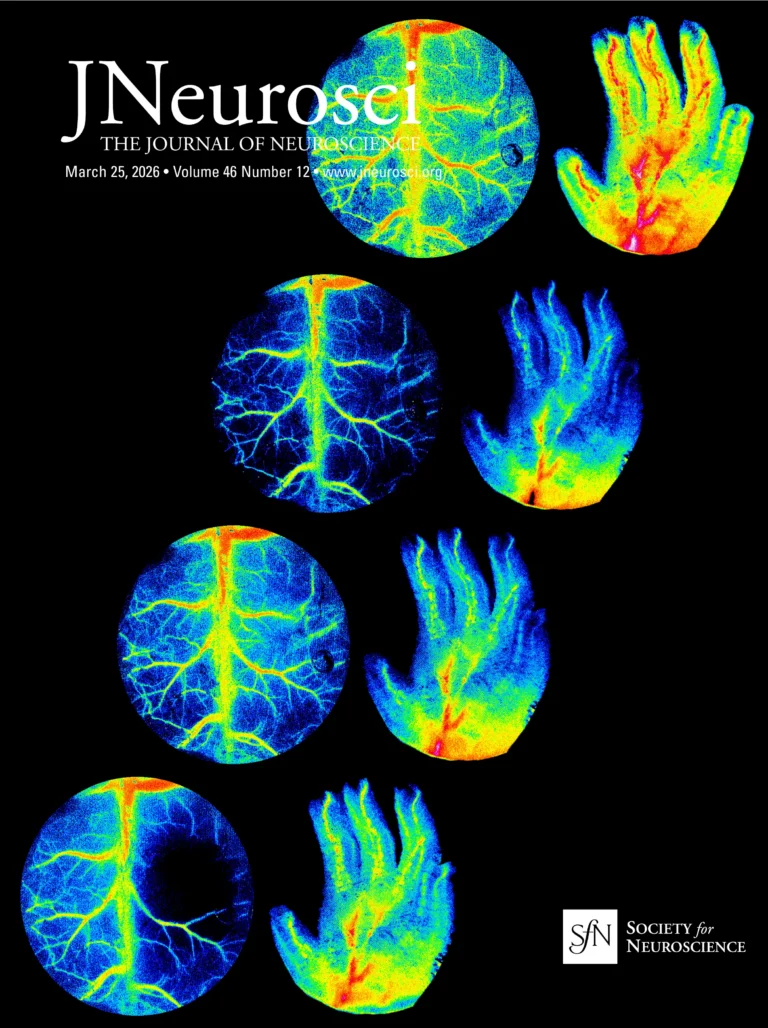
The Rose Bengal photothrombotic stroke model is widely used in preclinical neuroscience for its simplicity, minimal invasiveness, and precise spatial targeting of ischemia. Despite its