NiraWave™ M is a micellar formulation of the clinically established near-infrared dye indocyanine green. The optimized formulation enhances fluorescence performance, improves aqueous stability, and extends circulation time compared to free dye. These properties make NiraWave™ M suitable for optical angiography and for the visualization of vascular leakage associated with inflammatory processes.
By remaining within intact vasculature while extravasating at sites of increased permeability, NiraWave™ M enables sensitive detection of inflammation and microvascular alterations in preclinical models.
NiraWave™ M is well suited for:
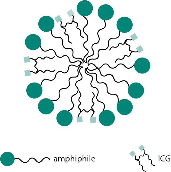
Micelle size: approximately 11 nm
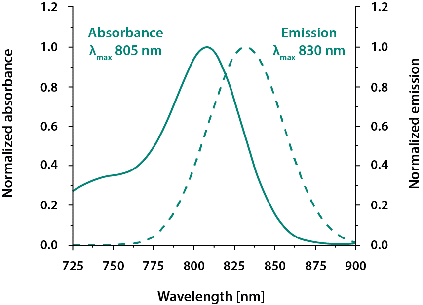
Emission wavelength: 830 nm
Excitation wavelength: 660 to 790 nm
The near-infrared spectral profile supports reduced background autofluorescence and improved tissue penetration in small animal imaging.
In optical angiography studies of the mouse ear, NiraWave™ M demonstrates extended circulation time compared to conventional indocyanine green. In inflammatory disease models, including rheumatoid arthritis in rats, increased vascular permeability leads to localized signal enhancement, enabling differentiation between inflamed and control tissue.
Normalized absorption and emission spectra in plasma confirm stable fluorescence characteristics suitable for quantitative imaging workflows.
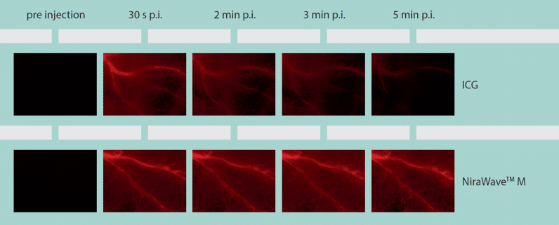

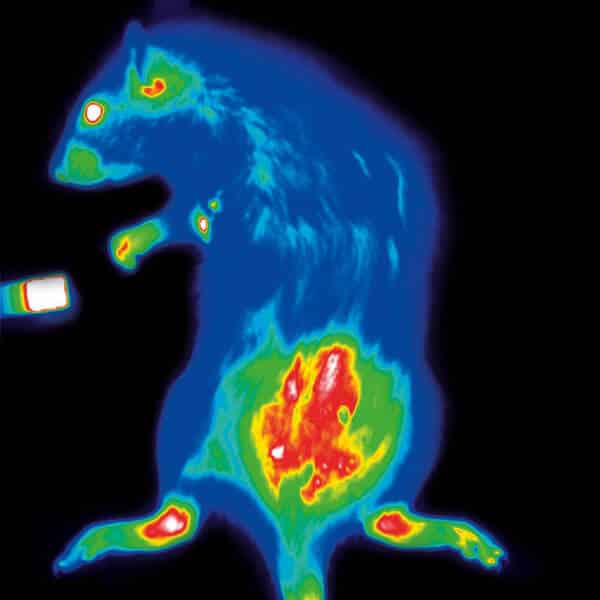
Top: Mouse ear optical angiography with NiraWave M shows superior circulation time over ICG standard dye.
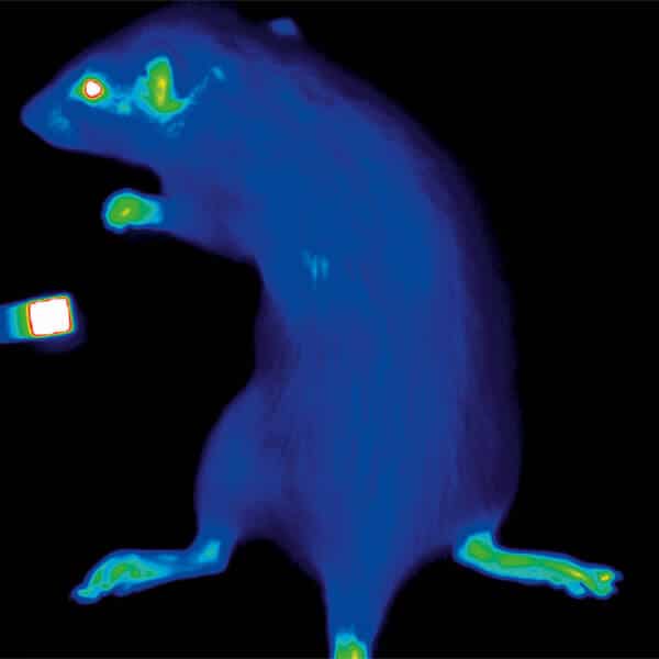
Bottom: Inflammation imaging in a rheumatoid arthritis rat model (right) versus the control animal (left) based on NiraWave M vascular leakage.
NiraWave™ M is available in single and multi-injection formats. Ordering through an authorized distributor ensures consistent product quality, technical support, and guidance for experimental design in preclinical optical imaging.
Comprehensive data sheets and technical documentation are available to support protocol development and study planning.
Authorized distributor of Viscover products by Nanopet Pharma GmbH. This page provides original distributor-specific content for preclinical inflammation imaging applications.