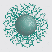
NiraWave™ Rocker is a nanoparticulate near-infrared (NIR) fluorescence imaging agent developed for sensitive detection of primary tumors and metastases in preclinical cancer models. Utilizing a rocker-switch uptake mechanism, the agent selectively accumulates in tumor cells, enabling high-contrast optical imaging with strong and stable fluorescence signals.
Its robust photophysical properties and tumor-selective enrichment make NiraWave™ Rocker a powerful tool for longitudinal oncology studies and therapeutic monitoring.
NiraWave™ Rocker is ideally suited for:

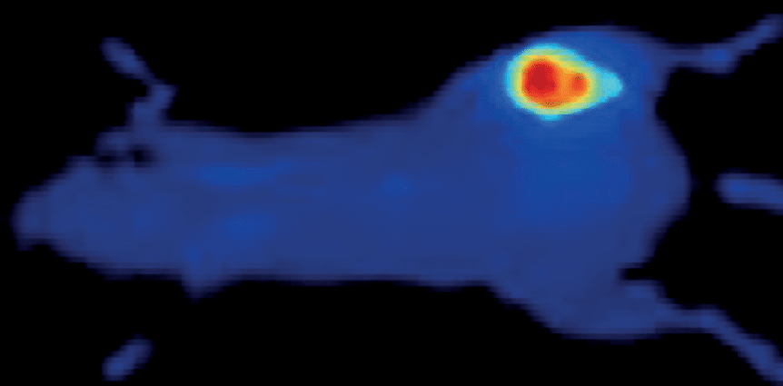
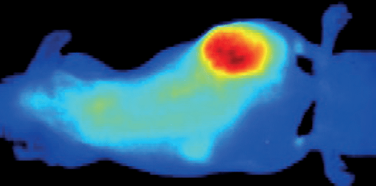
Top: Accumulation of NiraWave Rocker in tumors of a mouse model of human A549 lung adenocarcinoma (left) and murine T1 mammary carcinoma (right).
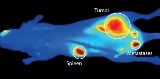
Bottom: Optical image of a mouse mopdel of human prostate cancer (PC-3) showing accumulation od NiraWave Rocker in primary tumor as well as in metastasized sciatic lymph nodes
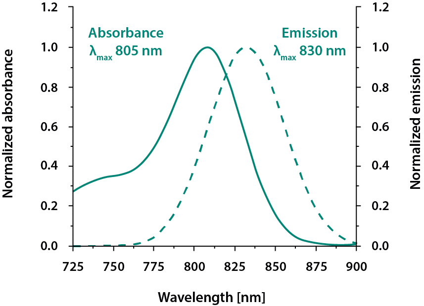
Emission wavelength: 830 nm
Excitation wavelength: 660–790 nm
The near-infrared spectral range supports deep tissue penetration and reduced background autofluorescence, enabling high tumor-to-background contrast in small animal imaging.
NiraWave™ Rocker demonstrates strong accumulation in multiple tumor models, including human A549 lung adenocarcinoma and murine T1 mammary carcinoma. Optical imaging studies further show clear signal enhancement in primary prostate cancer tumors (PC-3 model) and metastatic involvement of sciatic lymph nodes.
Normalized absorption and emission spectra confirm stable fluorescence behavior in plasma, supporting reproducible quantitative imaging.
NiraWave™ Rocker is available in single and multi-injection pack sizes. Ordering through an authorized distributor ensures reliable supply, technical guidance, and support for preclinical optical imaging studies.
Detailed data sheets and technical documentation are available to support experimental planning and imaging protocol optimization.
Authorized distributor of Viscover products by Nanopet Pharma GmbH. This page contains original distributor-specific content supporting preclinical tumor imaging using NiraWave™ Rocker.