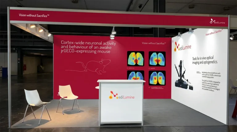
MediLumine at FENS 2026: Visit Us at Booth 159
We’re excited to be exhibiting at FENS 2026 in Barcelona, and we’d love to see you at Booth 159. Stop by to explore our full


Almost all animals with functional vision exhibit a variety of eye movements. These movements allow compensation for shifts in the visual scene and enable tracking of moving objects. Eye movements have been studied for over a century (Land 2006), and eye tracking has become an essential tool not only in neuroscience but also in fields such as neuromarketing (Gheorghe, Purcărea, and Gheorghe 2023). Additionally, eye tracking technology is increasingly integrated into virtual and augmented reality systems (Jin et al. 2024).
Eye movements are generated by three pairs of extraocular muscles that rotate the ocular globe within the orbit. Compared to limb movements, eye movements are simpler, making it easier to classify and quantify them.
Eye movements are commonly divided into gaze-stabilizing and gaze-shifting categories. Gaze-stabilizing movements compensate for motion of the visual scene or head, while gaze-shifting movements align the eyes with specific targets in the visual field.
Gaze-stabilizing movements include the optokinetic reflex (OKR), triggered by motion across large portions of the visual field, and the vestibulo-ocular reflex (VOR), which responds to head movements (Ambrad Giovannetti and Rancz 2024). Gaze-shifting movements include saccades, which are rapid shifts in gaze; vergence, which involves coordinated changes in the angle of both eyes; and smooth pursuit, which enables tracking of moving visual targets (Sekar, Panouillères, and Kaski 2024).
Understanding these movements has facilitated the use of eye tracking to study visual system mechanisms, motor control, and the early detection of neurodegenerative diseases such as Alzheimer’s and Parkinson’s, as abnormal eye movements often correlate with disease progression (Sekar, Panouillères, and Kaski 2024).
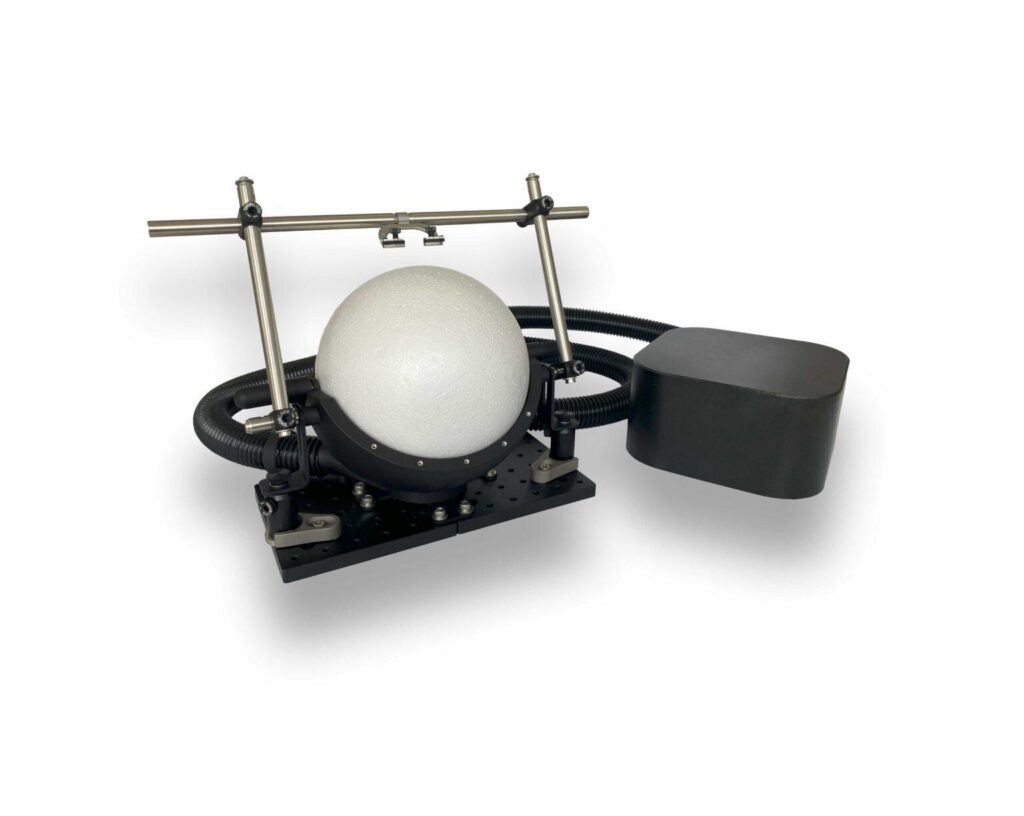
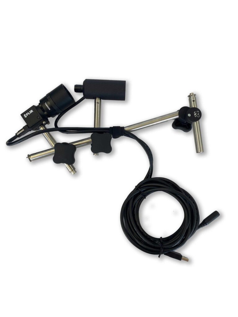
Several techniques exist to measure eye movements, including electro-oculography, magnetic scleral search-coils, and video-based eye tracking with infrared illumination. Video-based methods have become particularly popular in neuroscience due to their low invasiveness, high temporal resolution, and accessibility in both human and animal research.
Video-based eye tracking of a Human subject displaying a vestibulo ocular reflex (VOR). The acquisition was undertaken using the behavioral camera with infrared illumination and the pupil tracking was performed using the www.pupillometry.it website.
Mice are widely used as models to study brain structure and function. Eye tracking in mice allows researchers to assess visual performance, eye movement control, and overall brain state under various experimental conditions. For example, OKR measurements are commonly used to evaluate visual acuity in rodents.
The neural circuits controlling eye movements in mice are well characterized, involving the cortex, basal ganglia, superior colliculus, brainstem, and cerebellum (Stahl 2004). Eye tracking provides a quantifiable behavioral readout that can be used to evaluate pharmacological interventions or neurodegenerative disease models (de Jeu and De Zeeuw 2012; Betts-Henderson et al. 2010).
Video-based eye tracking also enables pupillometry, the measurement of pupil size dynamics. Pupil diameter reflects brain states, including alertness, attention, reward prediction, and even sleep stage (Yamada and Toda 2022). In rodents, pupil tracking combined with behavioral tasks provides an additional non-invasive tool to study cognitive and neural processes.
Eye tracking can be combined with imaging and behavioral analysis methods to yield comprehensive insights into visual processing and brain function. In mice, eye tracking data can complement widefield optical imaging and electrophysiology, providing a multidimensional view of neural activity.
Eye tracking is a versatile, cost-effective, and non-invasive tool for neuroscience research. Its applications in murine models enhance understanding of visual function, neural circuitry, and brain states, making it valuable for studies on neurodegenerative diseases and pharmacological interventions.
Ambrad Giovannetti E, Rancz E. Behind mouse eyes: The function and control of eye movements in mice. Neurosci Biobehav Rev. 2024;161:105671. doi:10.1016/j.neubiorev.2024.105671
Betts-Henderson J, Bartesaghi S, Crosier M, et al. The nystagmus-associated FRMD7 gene regulates neuronal outgrowth and development. Hum Mol Genet. 2010;19(2):342-351. doi:10.1093/hmg/ddp500
Gheorghe CM, Purcărea VL, Gheorghe IR. Using eye-tracking technology in Neuromarketing. Rom J Ophthalmol. 2023;67(1):2-6. doi:10.22336/rjo.2023.2
de Jeu M, De Zeeuw CI. Video-oculography in mice. J Vis Exp. 2012;(65):e3971. Published 2012 Jul 19. doi:10.3791/3971
Land MF. Eye movements and the control of actions in everyday life. Prog Retin Eye Res. 2006;25(3):296-324. doi:10.1016/j.preteyeres.2006.01.002
Sekar A, Panouillères MTN, Kaski D. Detecting Abnormal Eye Movements in Patients with Neurodegenerative Diseases – Current Insights. Eye Brain. 2024;16:3-16. Published 2024 Apr 9. doi:10.2147/EB.S384769
Stahl JS. Using eye movements to assess brain function in mice. Vision Res. 2004;44(28):3401-3410. doi:10.1016/j.visres.2004.09.011
Yamada K, Toda K. Pupillary dynamics of mice performing a Pavlovian delay conditioning task reflect reward-predictive signals. Front Syst Neurosci. 2022;16:1045764. Published 2022 Dec 8. doi:10.3389/fnsys.2022.1045764
*This article is licensed under a Creative Commons Attribution 4.0 International License, which permits use, sharing, adaptation, distribution and reproduction in any medium or format
We’re excited to be exhibiting at FENS 2026 in Barcelona, and we’d love to see you at Booth 159. Stop by to explore our full
The Rose Bengal photothrombotic stroke model is widely used in preclinical neuroscience for its simplicity, minimal invasiveness, and precise spatial targeting of ischemia. Despite its