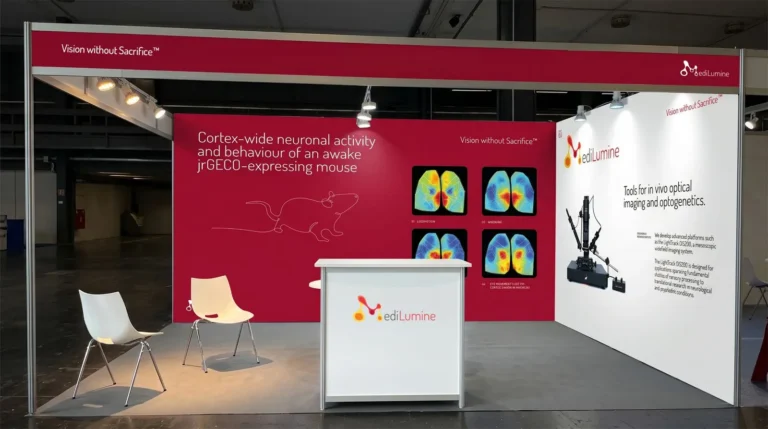
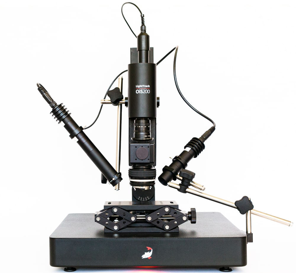
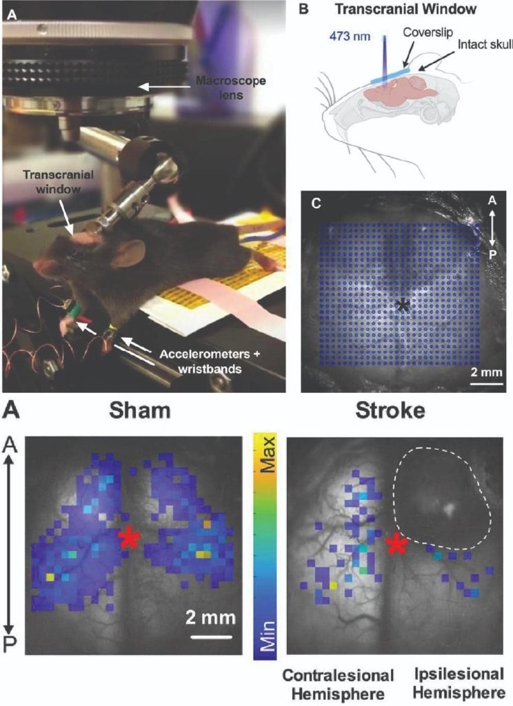
The LightTrack OiS200 Mesoscope for optical imaging and targeted optogenetics is a highly customizable system for in vivo experimentation. It enables scientist to simultaneously image calcium indicators, perform intrinsic optical imaging, laser speckle imaging for neurovascular coupling studies, and targeted optogenetics.
The integrated and ergonomic system allows you to save lab space and comes at an economical price point making it an ideal research companion for neuroscience, vascular biology and in vivo studies.
The technology allows for synchronization with many other types of equipment through user defined auxiliary and stimulation input/output options. Users can add two cameras and multiple illumination wavelengths allowing them to save the time required to build a customized rig.
Calcium Imaging
For calcium imaging, the LightTrack OiS200 system uses 475 nm laser based and LED based illumination (525 nm, 590 nm, and 625 nm) to obtain both calcium and intrinsic signals. The intrinsic signals are used to obtain the corrected calcium signal (ΔF/F, %) of each pixel. In the example image above, calcium imaging was performed with LightTrack OiS200 in head-fixed awake GCaMP6s adult mice before and after conditioning to understand long term plastic changes on cortical maps. ref. https://www.frontiersin.org/articles/10.3389/fncir.2020.00038/full
Cerebral Blood Flow and Hemodynamics
In this example, the LightTrack OiS200’s speckle imaging modality was used with 785 nm laser to image changes in blood flow and it’s intrinsic optical imaging modality was used for functional imaging of hemodynamics with intervals of 2 seconds and resolution at the micron scale. ref. https://www.frontiersin.org/articles/10.3389/fphys.2021.611984/full
Targeted Optogenetics
Targeted optogenetics can be performed using the laser stimulation matrix available on LightTrack OiS200 to generate bihemispheric motor maps. In this image, Thy1-ChR2-YFP mice having received a photothrombotic stroke at postnatal day 7 underwent logitudinal optogenetic motor mapping both before and after 3 weeks of skilled forelimb training. Optogenetic cortical stimulation enabled imaging of motor function biomarkers and showed that perinatal stroke reduces motor map size and movement latency AUC in both hemispheres. ref. ref. https://www.jneurosci.org/content/41/28/6157
Vascular Imaging
For vascular imaging, the LightTrack OiS200 uses the a 472 nm blue channel with the illumination and acquisition running at least 5 minutes. Other wavelengths are available upon request. In this example, fluorescent microbeads (FMOY-1.3, 460/594 nm, Cospheric, U.S.) were IV injected into C57/Bl6 mouse with scalp removed. The sum of all images was made in the analysis software showing the detailed anatomical structure of vasculature through the skull of the mouse.
Real time intrinsic optical imaging and laser speckle imaging of a mouse’s cortex following whisker stimulation. The mouse’s cortex was illuminated with interleaving light from red, amber, and green LED’s as well as a NIR laser . The images of cortex and fluorescent curves show hemoglobin (oxygenated, reduced and total) concentration and blood flow across the mouse’s barrel field cortex, a 2 x 3 mm ROI indicated by the blue dotted lines.
Contrast-enhanced in vivo microCT imaging is a powerful research method for enhancing soft tissue X-ray contrast, delineating soft tissue structures for PET or SPECT volume
We use cookies for various purposes including analytics and personalized marketing. By continuing to use the service, you agree to our use of cookies. - Cookie Policy