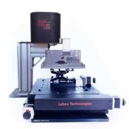
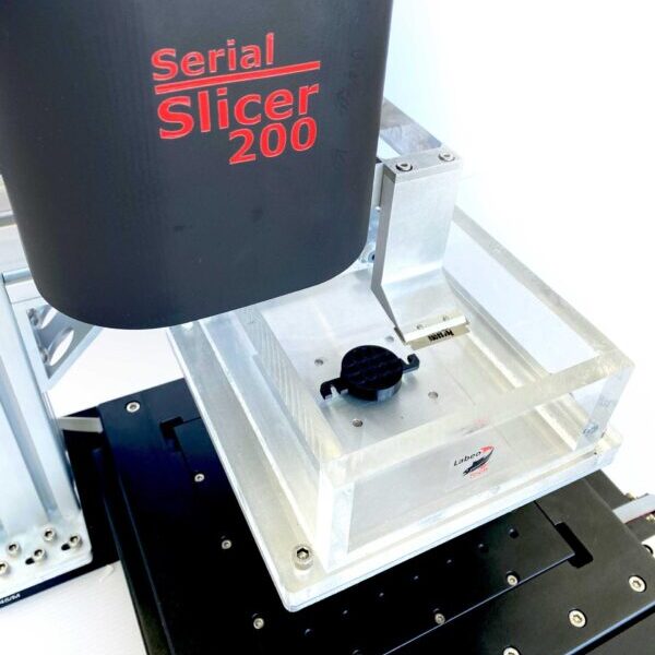
The Serial Blockface Histology Platform enables serialized three-dimensional histology of ex vivo tissues, such as rodent brains or small animal arteries. It is compatible with optical coherence tomography (OCT & PS-OCT), confocal fluorescence microscopy and many more. The apparatus is composed of a precise 3-axis motor kit to move a tissue sample fixed inside a water tank under a microscope (not included) for imaging in a mosaic pattern. A vibrating blade is then used to cut a thin layer of the tissue sample (typically 0.2 mm). This process is then repeated until the full depth of the tissue sample as been imaged.

With a fully integrated electronic and axis controller, the device requires no external components apart from the microscope, streamlining its setup and use.

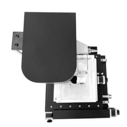
The motorized stage and slicer head feature a compact design, requiring minimal space and allowing for the use of external imaging systems. The device is also fully compatible with metric breadboards, such as optical tables, right out of the box.

The detachable water tub, constructed from high-quality solid cast acrylic, simplifies sample preparation and installation.
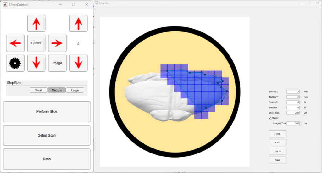
The software provides users with both automatic and manual control options, as well as the ability to select multiple regions of interest (ROI) for faster scanning. Additionally, users can load external images to aid in ROI identification. Setting up multiple scan parameters is also made easy with the software.
