
PRISM in vivo imaging systems now available through Kent Scientific
We are excited to announce that the PRISM In Vivo Imaging System is now available through Kent Scientific — makers of the SomnoFlo low-flow anesthesia


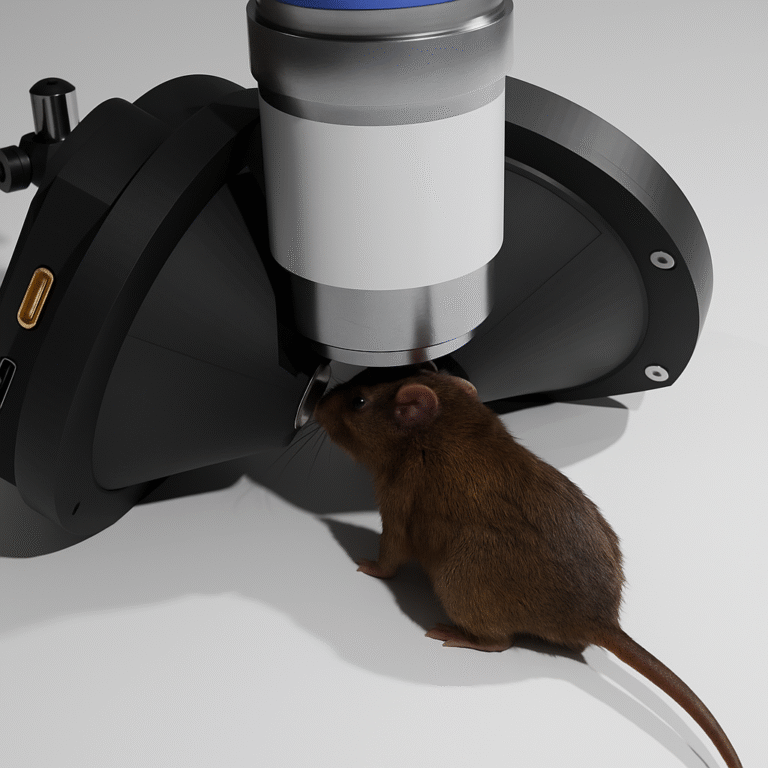
Investigating the neural mechanisms underlying behavior in awake, active animals remains one of the most pressing challenges in modern neuroscience. Conditions such as autism spectrum disorder, Alzheimer’s disease, and Parkinson’s disease all manifest as disruptions to behavior — making it essential that researchers study brain activity not in isolation, but in the context of real, ongoing actions.
The ideal scenario would be to record neural activity during unconstrained, naturalistic movement. In practice, however, this approach introduces substantial experimental noise. Natural behavior is rich and multisensory, involving constantly shifting stimuli and unpredictable movement trajectories — variables that are difficult to isolate or control. This limits the precision with which specific neural computations can be attributed to specific inputs.
Virtual reality (VR) has emerged as a compelling methodological solution. By presenting animals with dynamically responsive simulated environments, researchers can maintain behavioral engagement while retaining tight control over experimental variables. The result is a paradigm that bridges ecological validity and experimental rigor.
VR in neuroscience is not new. The technology has been applied to human subjects for decades, and its adoption in animal research has grown steadily — particularly in rodent studies of spatial navigation, memory encoding, and associative learning.
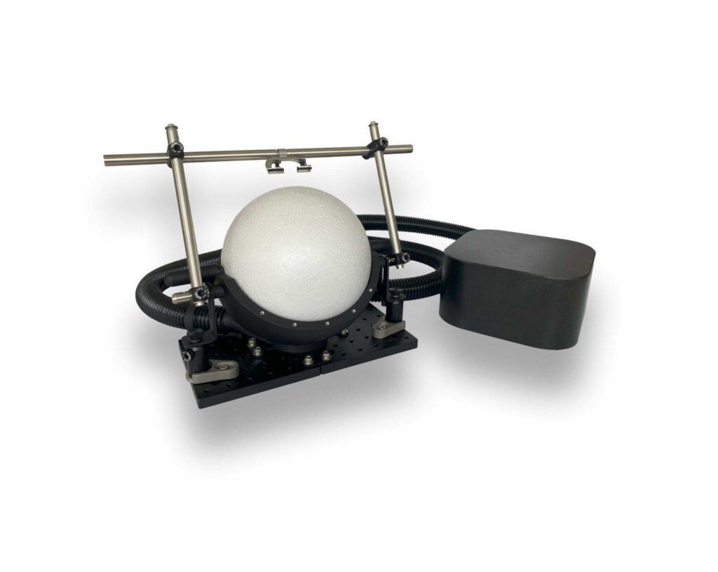
In head-fixed mouse preparations, VR is especially powerful. The animal runs on a spherical treadmill while visual (or multisensory) stimuli are projected around it, creating the experience of moving through a virtual world. Head fixation, which is required for most VR setups in rodents, simultaneously enables the use of recording modalities that demand mechanical stability — including widefield optical imaging, two-photon calcium imaging, and electrophysiology. This combination of behavioral engagement and recording access is difficult or impossible to achieve in freely moving animals.
VR environments can be tailored to target specific cognitive functions. Researchers have used them to study place cell dynamics, contextual fear conditioning, perceptual decision-making, task-switching, and more. The paradigm is highly flexible: sensory modalities, cue identities, task structure, and reward contingencies can all be adjusted to address specific experimental questions.
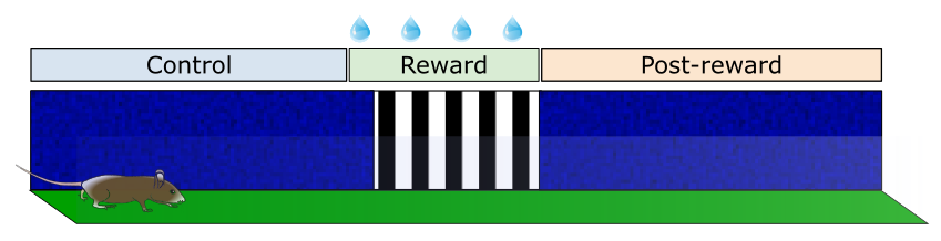
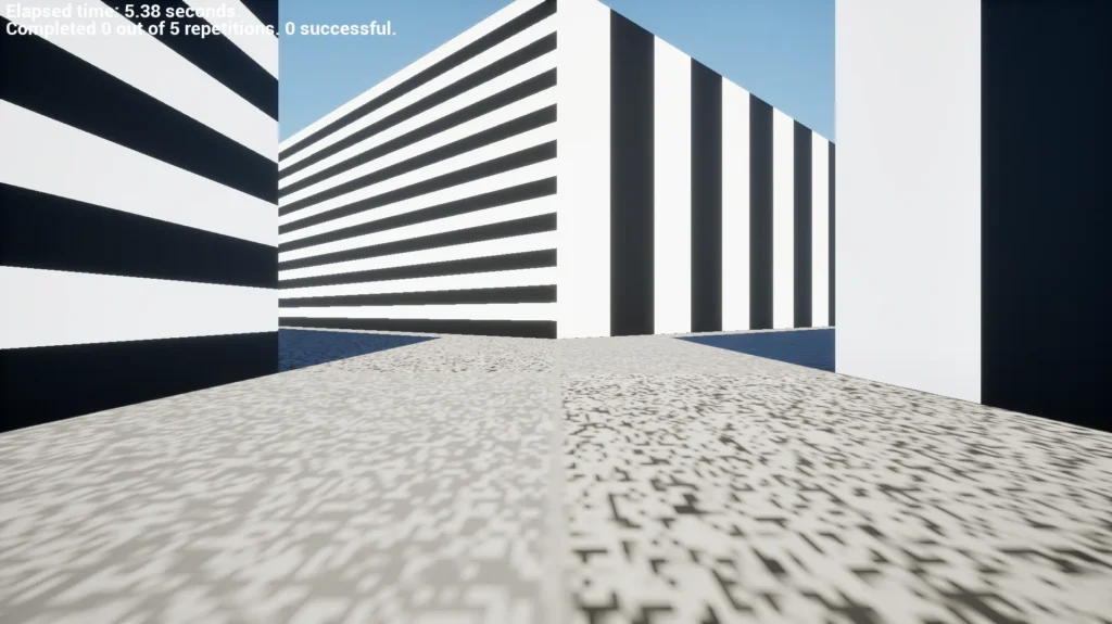
The experiment described here used a cohort of five head-fixed mice implanted with cranial headposts. Animals were positioned on a spherical treadmill and presented with a virtual environment through a dedicated mouse VR headset. The virtual track was 5 meters in length, with distinct wall patterns defining three functional regions:
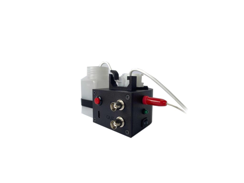
Liquid reward (sugar water) was delivered through a lick spout connected to a water dispenser equipped with a lickometer. Each session comprised 40 track traversals, with animals completing one session per day.
Training proceeded in two phases following an initial habituation period of five to seven days:
Pre-training (Days 1–3): Reward was delivered automatically upon entry into the reward zone, independent of licking behavior. This phase exposed animals to the contingency between the visual cue and reward without requiring an operant response.
Training (Days 4–9): Reward delivery became contingent on licking detected by the lickometer, and only within the reward zone. This phase required animals to actively engage with the reward zone based on learned cue-reward associations.
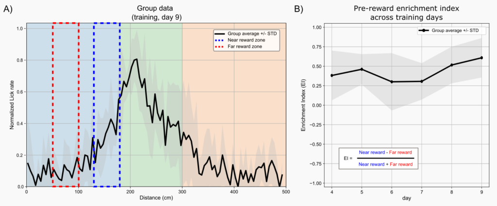
Lick rate was computed as the number of detected licks divided by time spent within 5 cm positional bins along the track. To facilitate group-level comparison, lick rate per trial was normalized to its maximum value.
A pre-reward enrichment index was derived as the normalized difference between the mean lick rate in the 50 cm window immediately preceding the reward zone and a baseline window located 100–150 cm upstream. Positive enrichment index values indicate elevated licking in anticipation of reward — a behavioral signature of learned cue-reward association.
All five animals developed robust anticipatory licking behavior across the training period, with some individuals displaying the pattern as early as day three. The behavioral shift was clear and consistent: whereas early sessions showed diffuse, exploratory licking distributed across the track, later sessions revealed a sharp increase in lick rate as animals approached the reward zone, followed by a return toward baseline in the post-reward zone.
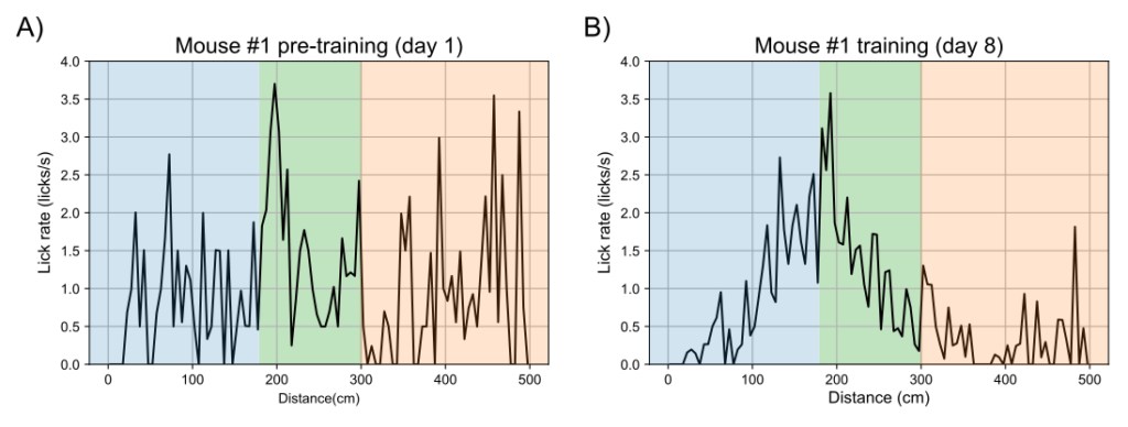
This spatial restructuring of licking behavior — peaking just before and during the reward zone — is a well-established index of associative learning. It indicates that the animal has formed a predictive representation linking the visual cue (vertical gratings) to the upcoming reward, and is acting on that prediction in advance of reward delivery.
By day nine, the group-level analysis confirmed that all five mice showed positive pre-reward enrichment indices, with values increasing progressively across training days. This trajectory is consistent with gradual consolidation of the learned association over repeated experience.
The findings from this experiment make a straightforward but important point: mice readily acquire goal-directed, cue-guided behavior in virtual environments. The learned anticipatory licking observed here is not a trivial response, it reflects the formation and expression of a predictive internal model, a process that depends on intact associative circuits spanning the striatum, prefrontal cortex, hippocampus, and dopaminergic midbrain systems.
From a methodological standpoint, this has several important implications.
Reproducibility and experimental control. Because the VR environment is fully programmable, every stimulus, spatial position, and temporal event can be reproduced exactly across animals and sessions. This degree of control is unattainable in open-field or unstructured environments, and it substantially reduces inter-trial variability.
Compatibility with brain recording. Head fixation, a prerequisite for most rodent VR setups, creates an ideal platform for concurrent neural recording. Calcium imaging, electrophysiology, and optical intrinsic signal imaging can all be performed while the animal is actively engaged in a learned task, enabling direct correlation between behavioral output and neural dynamics.
Translational relevance. The paradigm is well-suited for use in transgenic or lesion-based disease models. Deficits in cue-reward association, anticipatory behavior, or extinction learning are relevant to a wide range of neurological and psychiatric conditions, including addiction, depression, and neurodegenerative disease. VR-based tasks provide quantitative, repeatable measures of these functions that can be compared across experimental groups with high sensitivity.
This experiment demonstrates that virtual reality is an effective and versatile tool for probing learning and reward processing in head-fixed rodents. The rapid acquisition of anticipatory licking behavior — observed across all animals — confirms that mice engage meaningfully with virtual cues and form stable predictive associations within a naturalistic task structure.
As VR technology continues to improve in accessibility and resolution, and as its integration with neural recording modalities deepens, it is poised to become a core platform for systems neuroscience research. Its combination of behavioral control, recording compatibility, and ecological validity makes it uniquely suited to answering questions about how the brain learns, predicts, and responds to rewarding outcomes both in health and in disease.
We are excited to announce that the PRISM In Vivo Imaging System is now available through Kent Scientific — makers of the SomnoFlo low-flow anesthesia
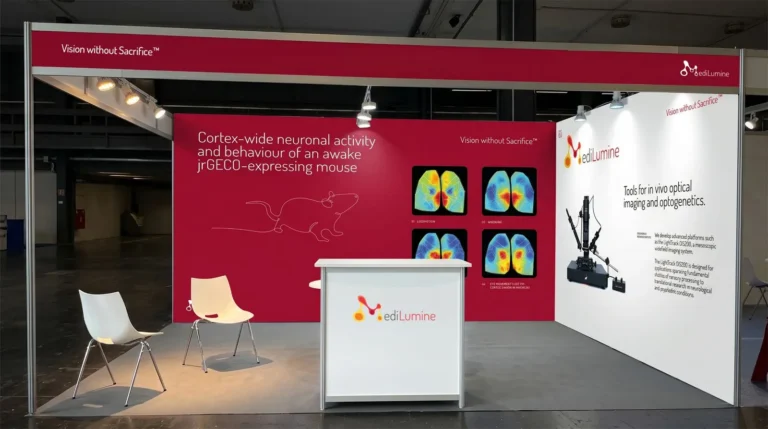
We’re excited to be exhibiting at FENS 2026 in Barcelona, and we’d love to see you at Booth 159. Stop by to explore our full