The Rose Bengal photothrombotic stroke model is widely used in preclinical neuroscience for its simplicity, minimal invasiveness, and precise spatial targeting of ischemia. Despite its popularity, the independent physiological effects of its two core components, the photosensitizer Rose Bengal (RB) and the activating light, had never been rigorously characterized in isolation. This gap matters: stroke outcomes are sensitive to even modest changes in blood flow, brain temperature, and large-scale electrical activity, meaning unaccounted physiological perturbations introduced by the model itself can confound experimental results.
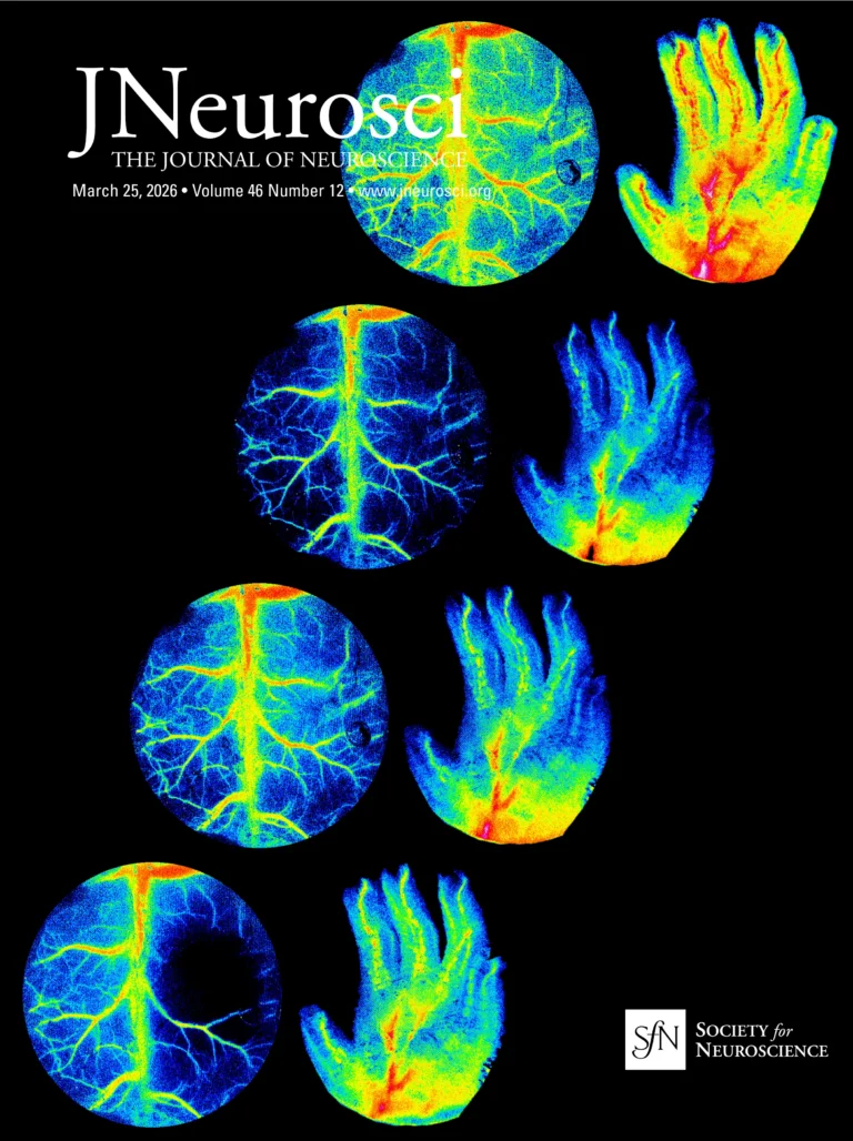
In a recent article published in the Journal of Neuroscience, Chary et al. (2026) provide the most comprehensive characterization of photothrombotic stroke induction to date. Using a multimodal approach combining laser Doppler flowmetry, laser speckle contrast imaging, functional ultrasound, two-photon microscopy, thermal imaging, and DC-EEG, including optogenetic stimulation performed with the LightTrack OIS200 Mesoscope, the authors systematically isolated the physiological contributions of each component of the photothrombosis protocol.
Rose Bengal is not physiologically inert
Even without photoactivation, Rose Bengal rapidly constricts cortical blood vessels and reduces blood flow throughout the brain and periphery, while also inducing transient bradycardia and tachypnea. These effects occur before any light is applied and represent a baseline physiological shift that all photothrombotic stroke experiments begin from.
The activating light alone causes significant tissue heating of approximately 3 degrees C and induces hyperemia measurable even in the contralateral hemisphere. Sham conditions that do not account for these effects separately from Rose Bengal introduce a source of confound that can distort experimental comparisons.
Yellow light at 561 nm, matched to Rose Bengali’s red-shifted absorption peak in plasma, produces a more pronounced and consistent drop in blood flow, earlier onset of spreading depolarizations, and significantly larger lesions than the commonly used green light at 520 nm. Importantly, 561 nm illumination does not cross-activate channelrhodopsin-2 (ChR2), making it compatible with optogenetic experiments.
Male mice developed consistently larger strokes than females across all cohorts. No intraoperative hemodynamic metric accounted for this difference, highlighting sex as a biological variable that must be considered in photothrombotic stroke study design and analysis.
Together, these findings reframe photothrombosis not as a simple on/off stroke switch, but as a sequence of competing physiological perturbations, each with independent consequences for experimental outcomes. Chary et al. provide clear, actionable guidance on sham design, light source selection, and the treatment of sex as a biological variable that will be valuable to any laboratory running photothrombotic stroke studies.
Source
Chary P, Rehmani S, Davidson S, Li X, Chen SX, Silasi G. Hemodynamic and Electrophysiological Progression of the Rose Bengal Photothrombotic Stroke Model in Mice. J Neurosci. 2026;46(12):e1818252026. Published 2026 Mar 25. doi:10.1523/JNEUROSCI.1818-25.2026