See Biology in True 3D with the HREM 3D Episcopic Imaging System
What if you could visualize your specimen in true 3D at micron-scale resolution, without tissue clearing, without alignment errors, and without the resolution limits of


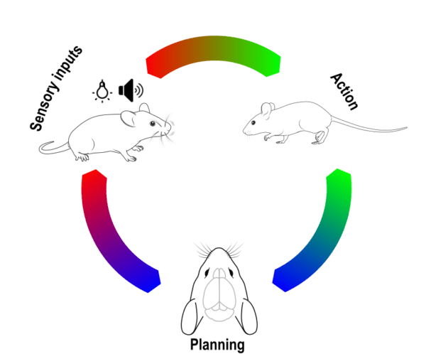
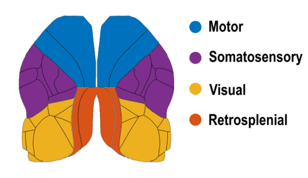
The mammalian cortex comprises multiple specialized regions that work in concert to perceive, plan, and execute behavior. In mice, as in other species, these regions are tightly integrated and dynamically adapt their interactions based on the behavioral context. Understanding how behavior modulates cortical dynamics is central to uncovering the brain’s organizational principles.
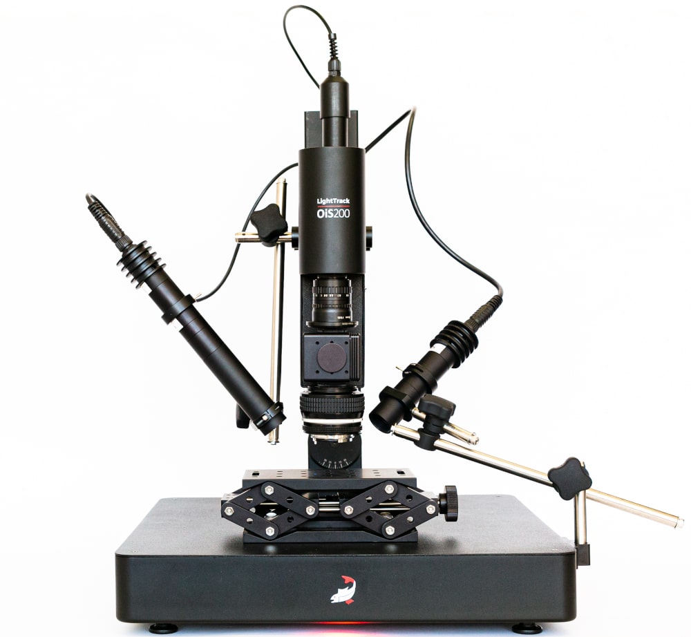
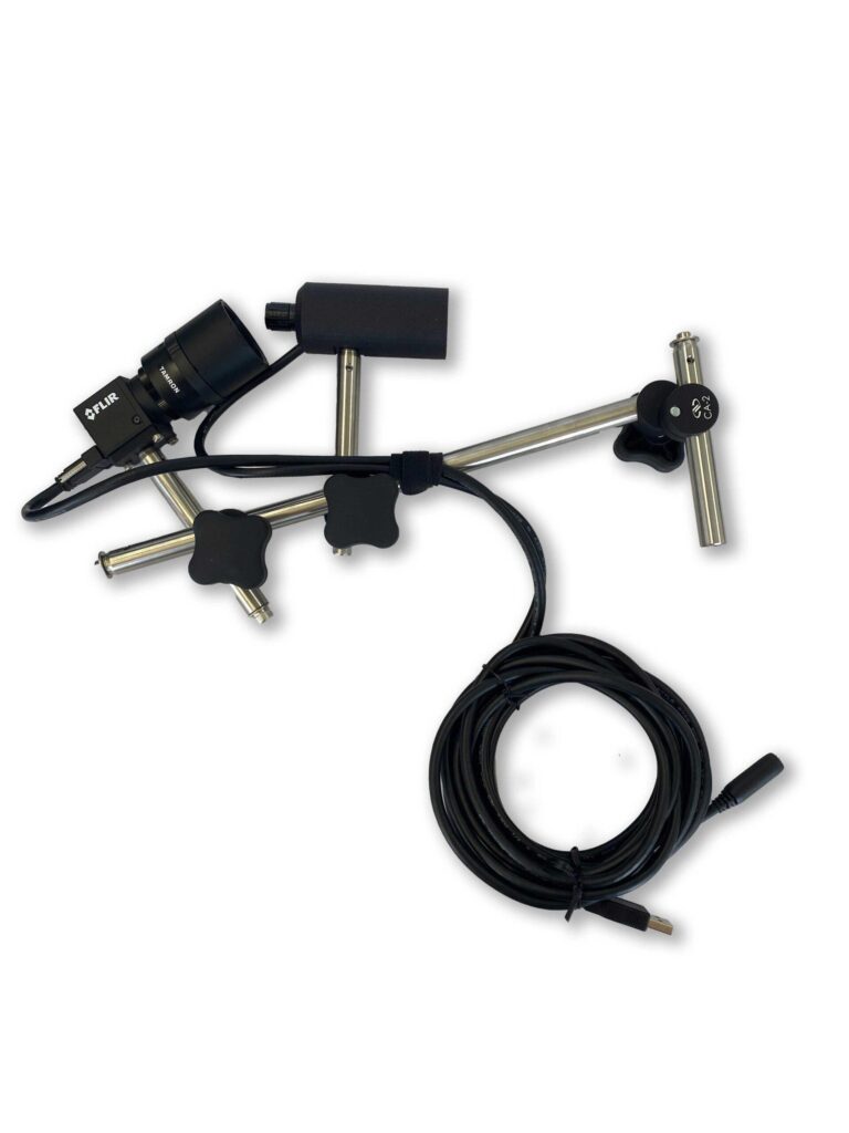
Widefield calcium imaging enables mesoscale recordings across the mouse neocortex. The high spatial coverage of this approach makes it well-suited to study how large-scale neural circuits interact during behavior. In this experiment, a transgenic mouse expressing the red calcium indicator jRGECO was imaged using the Light Track OiS200 system, with lime LED illumination at 567 nm and a frame rate of 20 Hz.
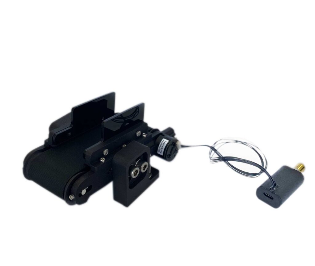
Spontaneous behavior was recorded for 30 minutes while the animal was awake and head-fixed on a low-friction belt treadmill. No explicit stimuli were applied. Behavior was concurrently captured via an infrared behavioral camera and the treadmill’s encoder signal.
Four primary behavioral states were identified:
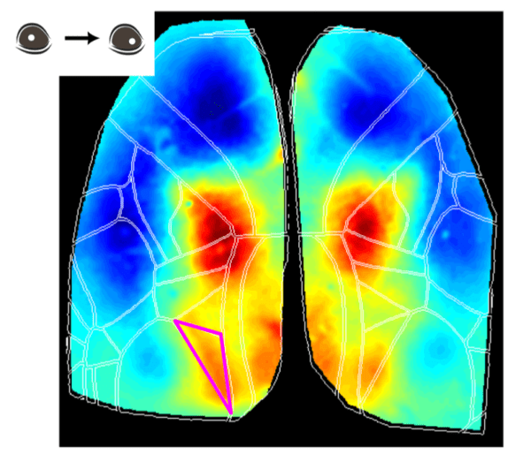
For non-locomotor behaviors, events were extracted using image-based features. For whisking and grooming, pixel intensity gradients within a region of interest (ROI) around the nose were used. Whisking was defined as changes >1 standard deviation (SD), grooming >2 SD. Eye movement was tracked by detecting shifts in pupil position, quantified as >2 SD displacement from baseline.
These behaviors were classified only during treadmill “quiet” periods to reduce confounding motor effects.

Fluorescence traces were processed via:
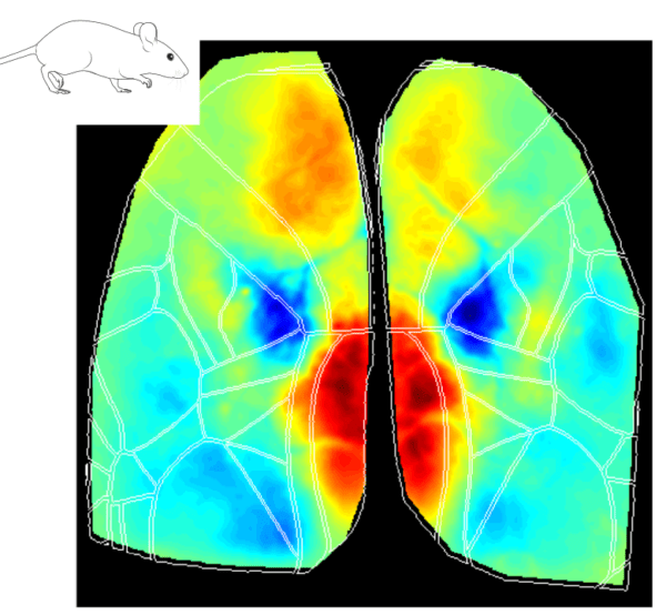
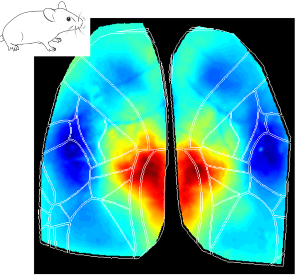
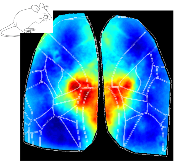
Each trial consisted of a 0.5s baseline and a 3s post-onset window. Activity was averaged across trials for each behavior, revealing consistent cortical activation maps.
Despite the spontaneity and partial overlap of some behaviors, clear spatiotemporal patterns emerged:
Across all behavioral states—locomotion, whisking, grooming, and eye movements—the Retrosplenial cortex (RS) showed early activation. RS has been implicated in spatial learning and head direction encoding (Vann et al. 2009; van der Goes et al. 2024), suggesting it plays a central role in contextual navigation, even in the absence of external stimulation.
These patterns reflect the neural substrates supporting spontaneous actions and suggest a modular yet integrated organization of the cortex.
This dataset demonstrates how widefield calcium imaging can uncover behaviorally relevant cortical dynamics in awake mice. Even in spontaneous, unstructured paradigms, consistent neural signatures align with specific behaviors. The robust activation of the retrosplenial cortex across conditions reinforces its proposed role in spatial cognition and internal mapping.
In the second part of this series, we will explore functional connectivity across cortical regions to investigate how networks reorganize in response to different behavioral states.
What if you could visualize your specimen in true 3D at micron-scale resolution, without tissue clearing, without alignment errors, and without the resolution limits of
Almost all animals with functional vision exhibit a variety of eye movements. These movements allow compensation for shifts in the visual scene and enable tracking