MediLumine at FENS 2026: Visit Us at Booth 159
We’re excited to be exhibiting at FENS 2026 in Barcelona, and we’d love to see you at Booth 159. Stop by to explore our full


We now turn our attention to interregional brain activity during periods of rest—providing additional insights into the brain’s intrinsic organization enabling studies aimed at assessing how such organization is altered in health and disease.
Functional connectivity refers to the temporal correlation between neural activities across different brain regions. These co-activation patterns offer valuable information about how the brain processes internal and external stimuli. In both healthy and pathological states, alterations in these patterns can indicate changes in brain function and structure. For example, disruptions in FC have been associated with neurodegenerative and psychiatric disorders such as Alzheimer’s disease and depression (Dennis & Thompson, 2014; Xu et al., 2024).

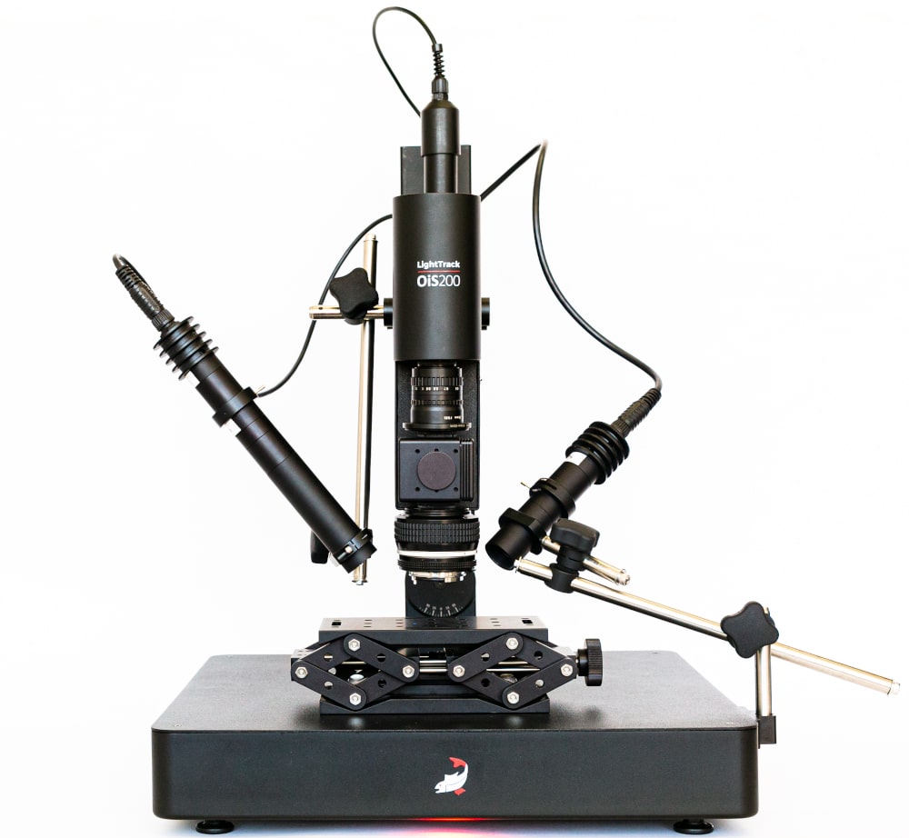
Resting-state functional connectivity (rs-FC) assesses how cortical regions interact in the absence of overt behavior or stimuli. The mouse neocortex, with its relatively accessible and well-characterized anatomy, is an ideal model for investigating rs-FC through widefield optical imaging. This approach has previously revealed functional network disruptions caused by stroke, hypoxia, and other neurological conditions (Bauer et al., 2014; Balbi et al., 2019; Bakker et al., 2023).
In this study, we built on prior behavioral analyses to isolate periods of rest in awake, head-fixed mice. Using a combination of behavioral video tracking and treadmill activity data, we identified intervals where the mouse was inactive. We then applied seed-pixel correlation analysis to fluorescence signals recorded via widefield imaging, after removing global oscillations to enhance signal specificity.
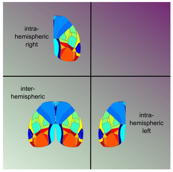
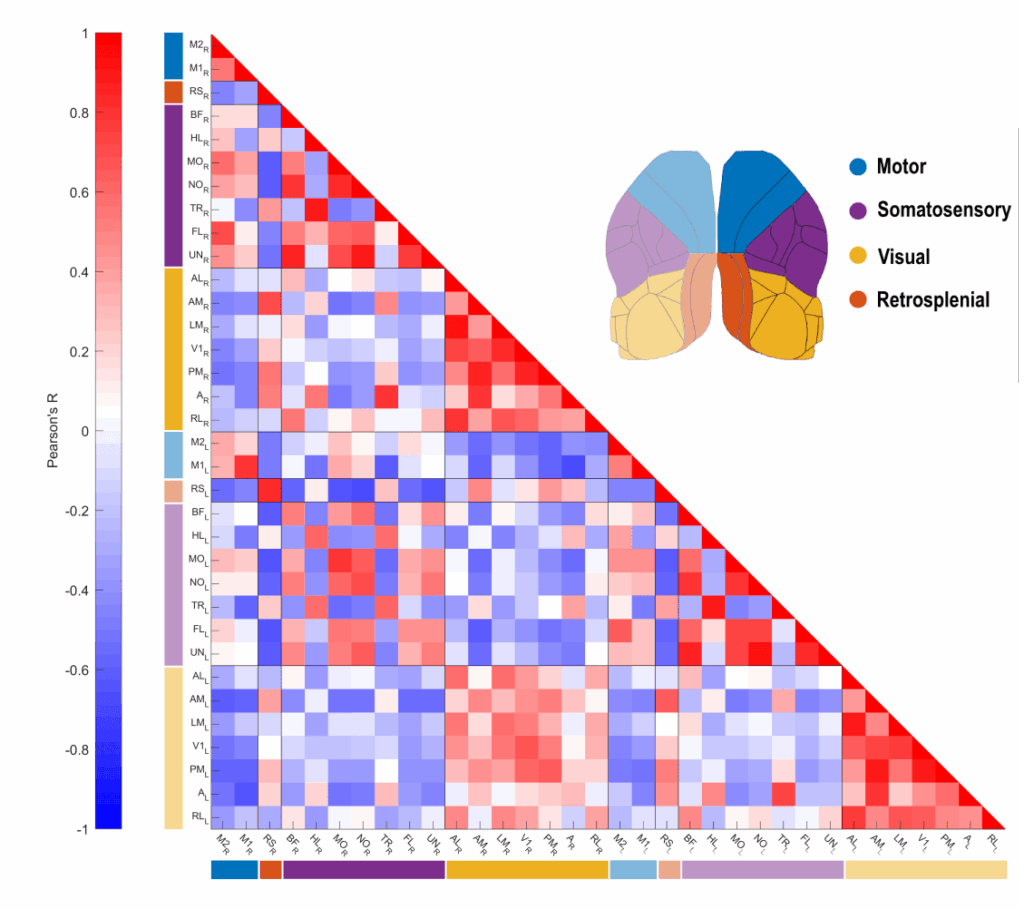
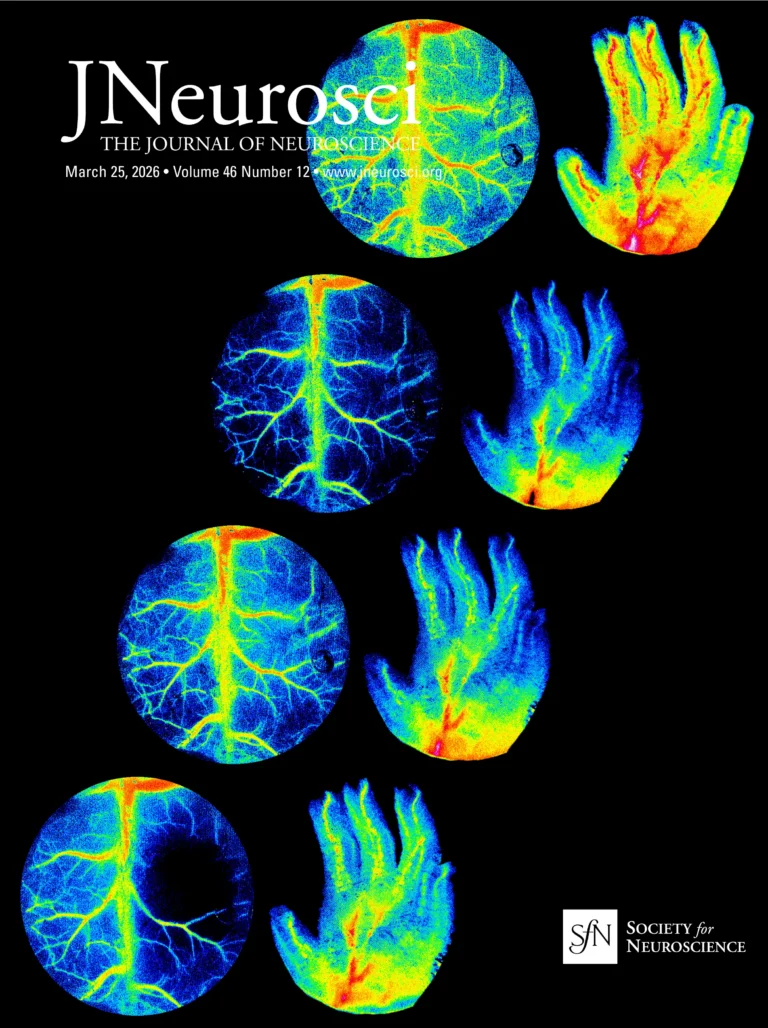
For each resting period, we calculated Pearson correlation coefficients between the averaged fluorescence signals of cortical areas. The resulting correlation matrix provides a clear, organized visualization of functional connectivity, with regions grouped by hemisphere and functional module. This format allows for intuitive comparisons between intra- and inter-hemispheric connectivity.
Our results are consistent with established resting-state connectivity patterns in mice. Intra-hemispheric correlations were generally stronger within functional modules—such as within motor or visual areas—than between them. Notably, we observed strong connectivity between motor and somatosensory regions, while visual areas showed higher correlation with the retrosplenial cortex. In contrast, visual and motor areas appeared largely anti-correlated, with weak correlations observed between visual and somatosensory areas.
This study demonstrates how widefield optical imaging can be used to investigate resting-state cortical activity in mice. By applying a straightforward experimental design, we were able to capture and characterize functional connectivity patterns reflective of the brain’s intrinsic organization. These findings underscore the value of widefield imaging in preclinical neuroscience, offering a robust platform for exploring how brain networks are altered in health and disease.
We’re excited to be exhibiting at FENS 2026 in Barcelona, and we’d love to see you at Booth 159. Stop by to explore our full
The Rose Bengal photothrombotic stroke model is widely used in preclinical neuroscience for its simplicity, minimal invasiveness, and precise spatial targeting of ischemia. Despite its