MediLumine at FENS 2026: Visit Us at Booth 159
We’re excited to be exhibiting at FENS 2026 in Barcelona, and we’d love to see you at Booth 159. Stop by to explore our full


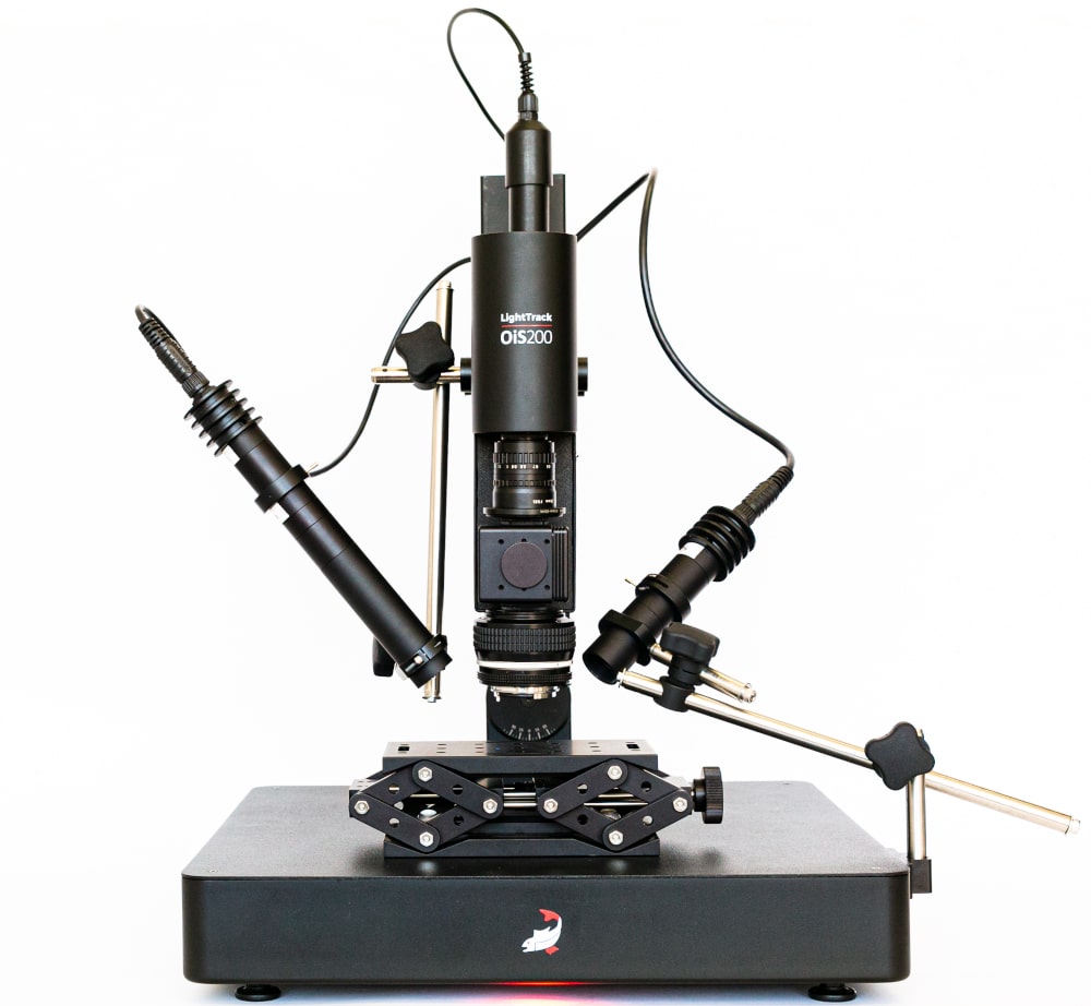
In previous articles, we introduced several approaches to study brain function using the LightTrack OiS200 platform, including intrinsic signals, autofluorescence, and calcium imaging. These techniques evaluate neuronal activity either directly, as in calcium imaging, or indirectly, through indicators such as metabolic expenditure or oxygen consumption.
Laser Speckle Contrast Imaging (LSCI) offers a complementary perspective. Instead of capturing neuronal activity itself, LSCI provides a visualization of vascular structure and blood flow in brain tissue. This technique enables the observation of cerebrovascular dynamics in real time, making it a valuable tool for investigating neurovascular interactions.
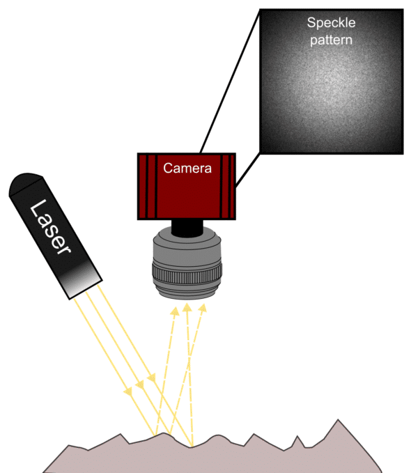
LSCI relies on the interference pattern generated when coherent laser light interacts with tissue. The reflected light, captured by a camera sensor, follows multiple optical paths within the tissue. The constructive and destructive interference of these light paths produces a random distribution of bright and dark regions, known as the speckle pattern.
In biological samples, moving particles such as red blood cells introduce fluctuations within the speckle pattern. When imaged over time, this motion causes a blurring effect that reflects the velocity of the underlying flow. Blood flow is therefore inferred by calculating the level of speckle blurring, quantified as speckle contrast, which is defined as the ratio of the standard deviation to the mean pixel intensity. Depending on the analytical approach, this calculation can be applied spatially or temporally, each method providing distinct advantages for specific applications.
Since its introduction in the 1980s, LSCI has been applied to visualize blood flow in various tissues. In neuroscience, it has become an important method for examining the vascular component of brain function. When combined with imaging modalities such as intrinsic signal or calcium imaging, LSCI enables a more complete characterization of neurovascular coupling and its pathological alterations.
Several studies have demonstrated its value in exploring disease-related impairments of vascular regulation. For example, LSCI has been employed to assess altered neurovascular coupling in mouse models of Alzheimer’s disease, as well as to investigate cerebral perfusion changes in traumatic brain injury and stroke models.
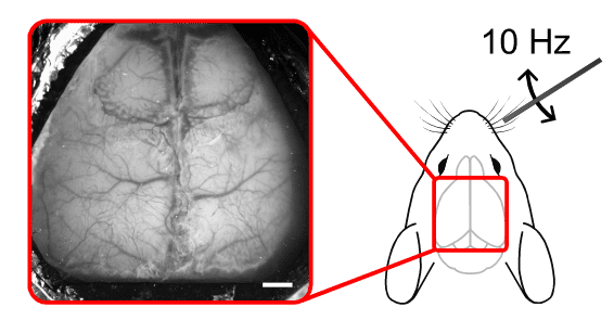
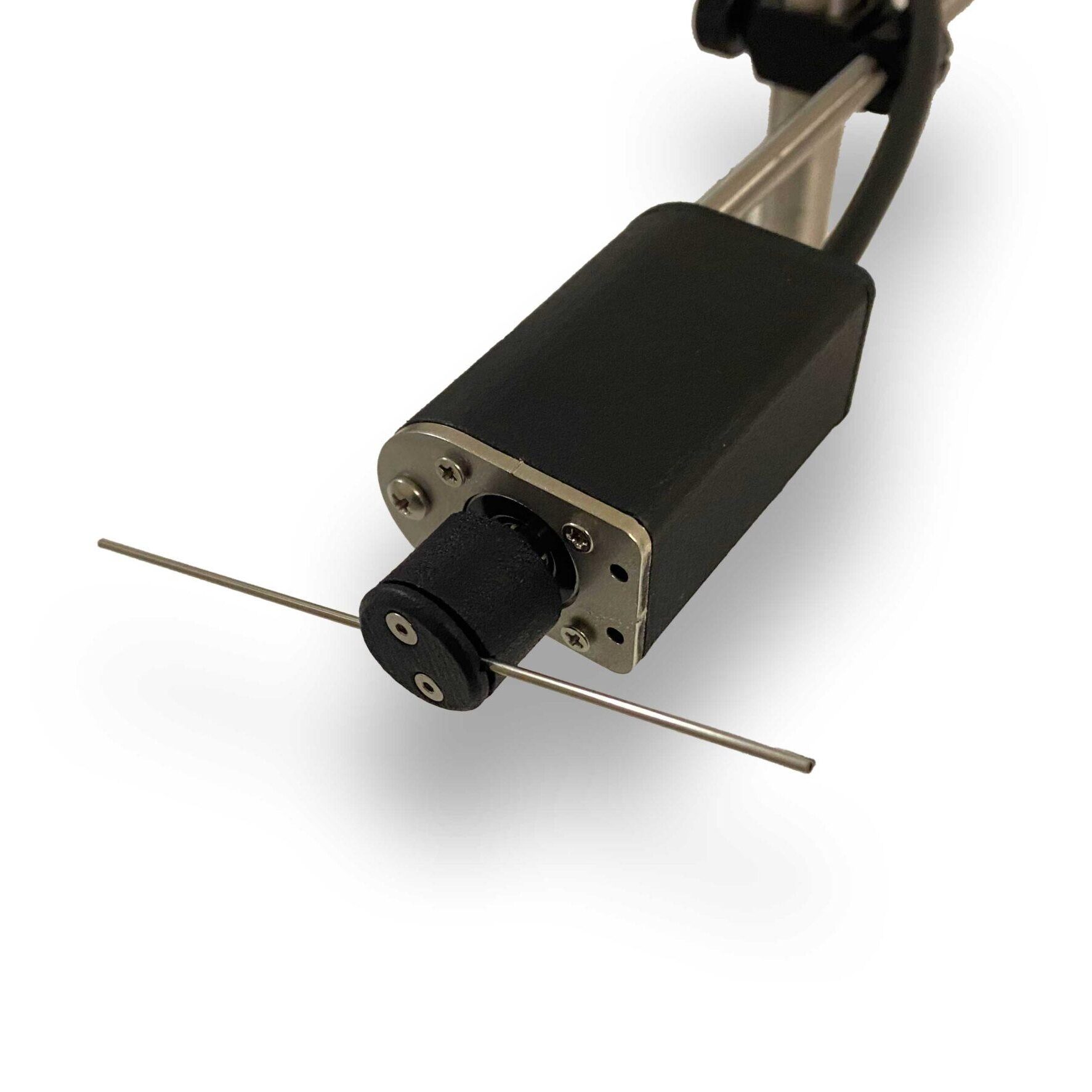
In a representative experiment, cortical responses were recorded in the left barrel cortex of a wild-type mouse under urethane anesthesia. Whisker stimulation was applied to the right whisker pad using a metal rod connected to a Mechanical Whisker Stimulator. The stimulation protocol consisted of ten-hertz oscillations in the horizontal plane for five seconds, followed by a twenty-second inter-stimulus interval. Each trial was repeated ten times.
Speckle data were acquired using a 785-nanometer laser and captured at thirty frames per second with a five-millisecond exposure time on the OiS200 LightTrack Imaging System. Anatomical reference images were recorded under green illumination.
Data analysis was performed using umIT, the Universal Mesoscale Imaging Toolbox, which provides a graphical user interface for processing large imaging datasets. The open-source software, available on the Labeo Technologies GitHub repository, includes detailed documentation and tutorials to facilitate data exploration.
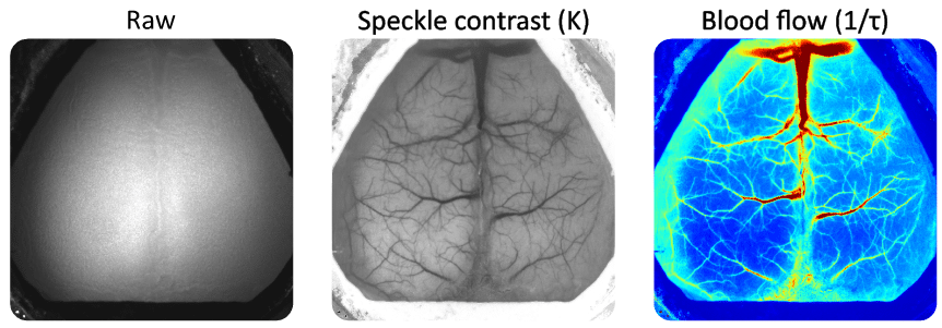
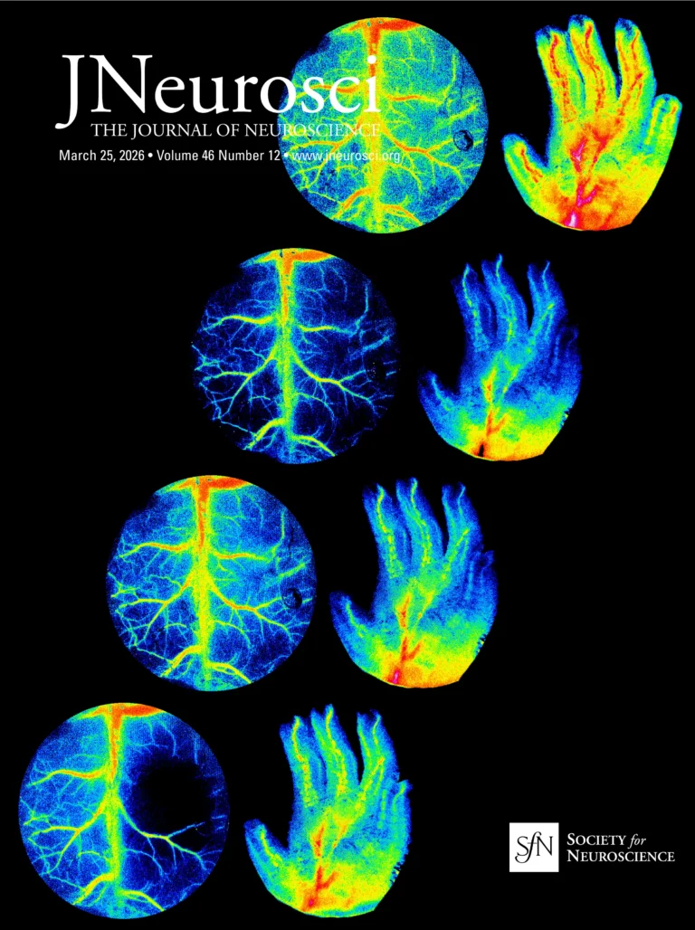
Raw speckle images were converted into speckle contrast maps and then processed to infer cerebral blood flow using standard computational algorithms. Data were temporally aligned, normalized to pre-stimulus baselines, and spatially filtered to enhance signal quality. The processed maps revealed localized increases in blood flow corresponding to whisker stimulation, confirming the link between sensory input and vascular response in the somatosensory cortex.
Analysis of the averaged trials showed a transient increase in blood flow within the left somatosensory cortex following contralateral whisker stimulation. The observed changes were temporally aligned with the stimulation period, demonstrating the effectiveness of LSCI in detecting rapid hemodynamic responses to sensory events.
Laser Speckle Contrast Imaging provides an accessible and informative method for studying cerebral blood flow. Its ability to visualize vascular dynamics without the need for exogenous dyes or contrast agents makes it a versatile addition to multimodal imaging workflows. When used in conjunction with intrinsic and calcium imaging, LSCI supports a comprehensive understanding of neurovascular coupling and cerebrovascular regulation.
This experiment highlights the potential of LSCI, combined with tools such as the Mechanical Whisker Stimulator and the OiS200 Imaging System, to advance research on sensory processing and cerebrovascular function.
We’re excited to be exhibiting at FENS 2026 in Barcelona, and we’d love to see you at Booth 159. Stop by to explore our full
The Rose Bengal photothrombotic stroke model is widely used in preclinical neuroscience for its simplicity, minimal invasiveness, and precise spatial targeting of ischemia. Despite its