Introducing the Rvein™: Easier Rat Tail Vein Injections, Starting Now
Anyone who has performed tail vein injections knows the challenge. The animal needs to be secure, the tail needs to be accessible, and the whole


What if you could visualize your specimen in true 3D at micron-scale resolution, without tissue clearing, without alignment errors, and without the resolution limits of conventional imaging?
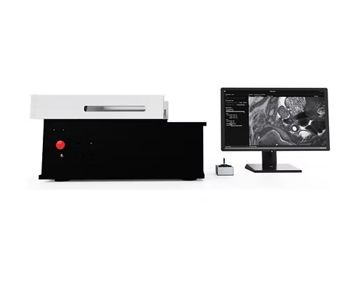
The HREM 3D Episcopic Imaging System, developed by Indigo Scientific, represents a fundamental shift in how researchers approach 3D imaging. This is not just another upgrade. It is a new standard for capturing biological structures with clarity, precision, and context.
For years, researchers have had to compromise when choosing imaging methods.
MicroCT offers whole-sample imaging, but struggles with soft tissue contrast and cannot achieve micrometer-scale resolution across large specimens. Meanwhile, Confocal Microscopy and Light-Sheet Microscopy require tissue clearing, which is time-intensive and can introduce artifacts, especially in dense or calcified tissues.
Even Histology, the gold standard for resolution, comes at a cost. Physical sectioning disrupts 3D structure, often leading to distortion, misalignment, and loss of spatial context.
The result is a persistent trade-off between resolution and anatomical integrity.
The HREM 3D Episcopic Imaging System eliminates this compromise through High-Resolution Episcopic Microscopy.
Instead of imaging thin sections individually, HREM captures images directly from the blockface of a resin-embedded specimen during automated sectioning. Each image is inherently aligned, producing perfectly registered stacks that reconstruct into accurate, distortion-free 3D volumes.
No clearing. No post-processing alignment. No loss of context.
The impact of HREM is already being felt across a wide range of disciplines.
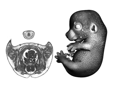
Capture the full 3D architecture of mouse embryos with histological clarity. From whole-body phenotyping to detailed organ morphometry, HREM preserves both resolution and spatial context.
As the field moves toward New Approach Methodologies, HREM offers a powerful tool for 3D characterization of organoids and other in vitro models. Capture internal architecture, lumen formation, and cellular organization at micron-scale resolution — without sectioning artifacts or clearing-induced distortion — making HREM ideal for validating and advancing NAM-based workflows.
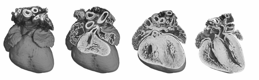
Visualize brain structures, connectivity, and fine morphology at a level that traditional imaging modalities cannot achieve. HREM bridges the gap between macro-scale imaging and cellular detail.
Screen structural phenotypes across cohorts with quantitative 3D morphometrics, even in dense tissues that previously required complex and time-consuming preparation.
Watch how the HREM 3D Episcopic Imaging System captures a complete 3D mouse embryo with exceptional clarity and detail.
Anyone who has performed tail vein injections knows the challenge. The animal needs to be secure, the tail needs to be accessible, and the whole
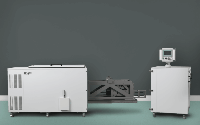
Bright Instruments is a UK-based manufacturer with more than 80 years of heritage in precision cryostat and microtome manufacturing. Trusted by pharmaceutical companies, CROs, and