MediLumine at FENS 2026: Visit Us at Booth 159
We’re excited to be exhibiting at FENS 2026 in Barcelona, and we’d love to see you at Booth 159. Stop by to explore our full


Vascular structure of the tumour. Three-dimensional rendering of the vascular structure of the LM8 osteosarcoma tumour as visualized in μCT after staining with Vascupaint.
Charged particle therapy with protons and carbon ions has become a central strategy in radiotherapy because of its favourable depth–dose distribution (the Bragg peak). Yet, clinical implementation faces a persistent challenge: range uncertainty. Even millimetre-scale errors in dose deposition can expose adjacent critical structures to unintended radiation.
In their recent Nature Physics article, Boscolo et al. (2025) report a preclinical study that addresses this limitation using radioactive ion beams (RIBs). By irradiating osteosarcoma-bearing mice with 11^{11}11C-ion beams and simultaneously acquiring in-beam PET images, the team demonstrated real-time beam range verification and full tumour control at high doses (20 Gy). Importantly, the study also revealed dose-dependent biological washout kinetics of the PET signal, suggesting a vascular component underlying tracer clearance.
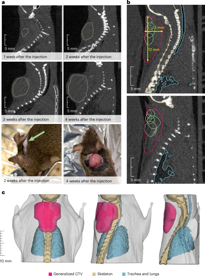
To validate these functional findings, the researchers turned to Vascupaint. Quantitative data such as radioactive washout curves provide valuable information, but they can sometimes leave critical questions unanswered. Do these measurements truly reflect biological processes, or are they influenced by decay artefacts? This is where Vascupaint served as what the authors call a “biological reality check.”
The integration of functional imaging (PET) and structural imaging (microCT with Vascupaint) represents a methodological advance with broader implications for preclinical oncology:
In the context of Boscolo et al.’s groundbreaking demonstration of radioactive ion beam therapy in osteosarcoma models, Vascupaint played a pivotal supporting role. By perfusing tumours and enabling 3D vascular reconstructions with microCT, it confirmed that PET washout dynamics reflected dense vascularization rather than experimental artefact.
Thus, Vascupaint emerges not only as a tool for vascular mapping, but also as a biological validation method that bridges the gap between functional measurements and anatomical reality. For researchers navigating the complexities of tumour biology, this dual role is invaluable: it ensures that experimental results are not only measurable but also make biological sense.
Boscolo, D., Lovatti, G., Sokol, O. et al. Image-guided treatment of mouse tumours with radioactive ion beams. Nat. Phys. (2025). https://doi.org/10.1038/s41567-025-02993-8
*This article is licensed under a Creative Commons Attribution 4.0 International License, which permits use, sharing, adaptation, distribution and reproduction in any medium or format
We’re excited to be exhibiting at FENS 2026 in Barcelona, and we’d love to see you at Booth 159. Stop by to explore our full
The Rose Bengal photothrombotic stroke model is widely used in preclinical neuroscience for its simplicity, minimal invasiveness, and precise spatial targeting of ischemia. Despite its