Advances in In Vivo Bioluminescence Imaging
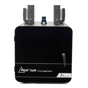
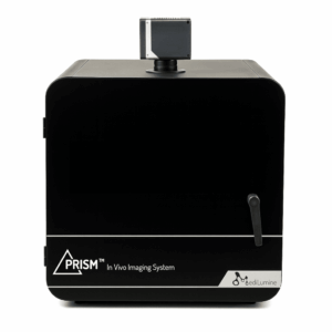
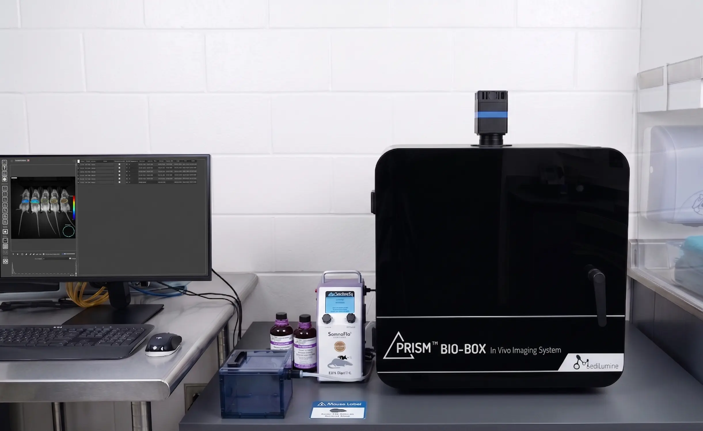
The PRISM™ BIO-BOX for five or 10 mouse in vivo imaging has the best price to performance ratio on the market today with a field of view of 22.7 cm. In BLI, what decides whether you see your signal is not raw sensitivity but contrast, the ratio of the bioluminescent signal to the background it sits against. A detector that collects a large photon flux gives no advantage if it also generates a large background, because the signal is swallowed by the noise. Real luciferase tumors are bright, well above one hundred thousand photons/s/cm²/sr, so the goal is not exotic hardware but a system that renders those signals cleanly against a quiet floor. That is exactly what the PRISM BIO-BOX delivers. Powered by the new SONY IMX428LLJ sensor, it is a highly economical solution for imaging bioluminescent signals, weak or strong, at a fraction of the cost of sensitivity you would never put to work.
PYRAMID Analysis Software
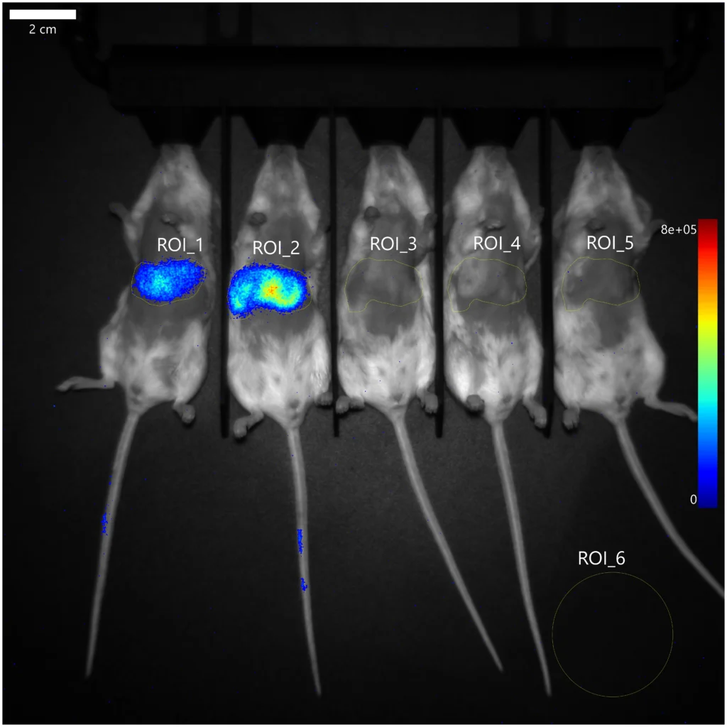
Pyramid analysis software is an intuitive imaging software designed to help you visualize, explore, and analyze your experimental data with ease. It has been optimized to work with data from the PRISM imaging platform, including bioluminescence, fluorescence and SWIR modalities. Pyramid lets you load multiple images, draw and adjust regions of interest (ROIs), and instantly see key statistics.
Applications
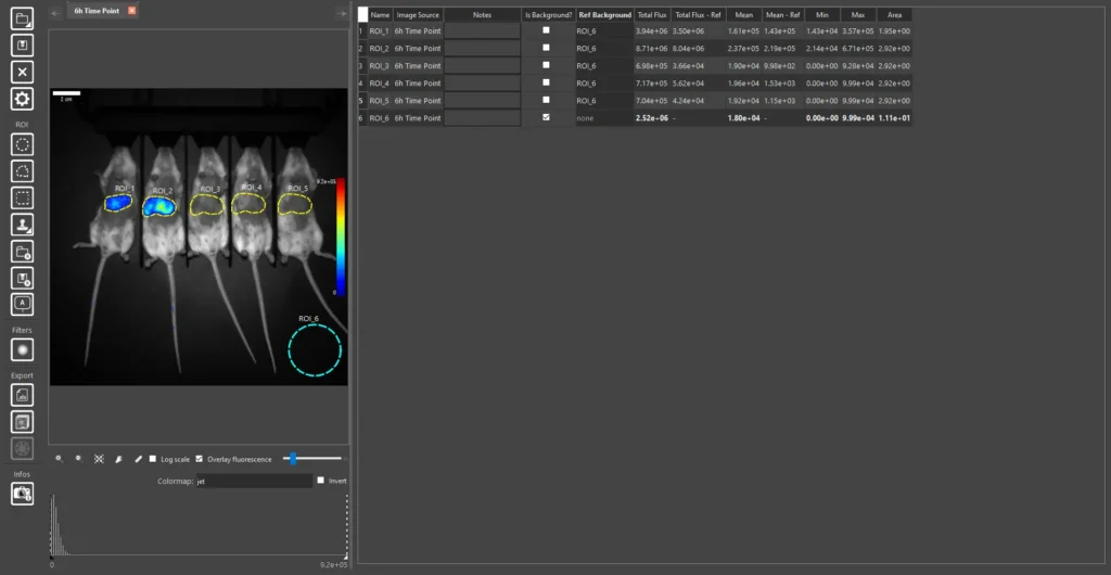
The PRISM BIO-BOX is a bioluminescence imaging system built for the clean detection of low-light signals across a range of preclinical and in vitro work. It is well suited to imaging autoluminescent bacteria in microbiology studies, monitoring tumor development in mice, exploring light emitting pathways in plants, performing cell culture imaging, and serving as a flexible alternative to traditional plate readers for luminescence based assays. Because the system pairs high photon detection with a low background, it supports researchers working with weak bioluminescent signals in both live animal and in vitro models.
Resolution is not sacrificed for sensitivity. The cooled CMOS sensor delivers a 3216 pixel wide array, roughly 7 megapixels, so faint sources are rendered as sharp, well localized spots rather than soft blooms. Cooling holds the dark background low through the long exposures that low-light imaging requires.
The platform is also expandable. An optional broadband light source with motorized filter wheels for excitation and emission filters adds true fluorescence imaging alongside bioluminescence, so a single instrument covers fluorescent reporters and dyes as well as luminescent signals, without the cost or footprint of a dedicated fluorescence system. A variable stage adjusts the field of view from 4 cm for single mouse imaging up to 20 cm for imaging five mice at once, so the same system scales from single subject studies to higher throughput cohorts.
SomnoFlo Anesthesia
All PRISM in vivo imaging systems come standard with an integrated Kent Scientific SomnoFlo anesthesia system. Unlike conventional setups, SomnoFlo requires no oxygen concentrator, oxygen tank, or house gas line; it draws and delivers ambient air directly, eliminating the need for a separate compressed gas supply chain. The advantage of working with SomnoFlo is simplicity and independence: your imaging suite doesn’t need to be built around gas infrastructure, permitting, or tank management, there’s no O2 supply to monitor, refill, or service, and there’s no risk of an imaging session being interrupted by a depleted tank or concentrator downtime, so labs and facilities without existing oxygen infrastructure get a fully self-contained, plug-and-play imaging and anesthesia workflow from day one.